The transvenous and epicardial operations are the two main
techniques of pacemaker implantation. The transvenous method involves
the insertion of an electrode lead into a vein, found at the shoulder or the
root of the neck. The lead is negotiated, under X-ray vision, into the correct
chamber of the heart and wedged or secured in position. The quantity of
electrical energy needed to stimulate contraction of the heart is tested. If this
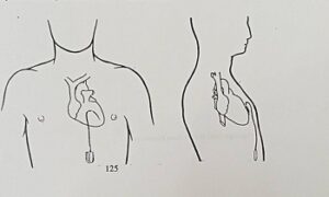
is satisfactory the electrode lead is connected to the pulse generator which
is then fitted into a small pocket fashioned between the skin and muscle of
the chest (Fig. 7). Modern pacemakers are so small that they are not
prominent and are usually almost hidden by overlying tissue. This
implantation method takes about half to one hour and is usually performed
under local anaesthetic without pain or discomfort to the patient. The
majority of pacemakers are inserted by the transvenous route.
Alternatively the pacemaker electrode lead can be attached or sewn
directly onto the outer surface, or “epicardium” of the heart. Pacemakers
attached to such “epicardial” electrode leads are usually sited in the tissues
of the abdominal wall (Fig. 8). The epicardial method is the preferred
treatment for patients undergoing other forms of heart surgery. ![]() After implantation of a pacemaker system by the transvenous route the
After implantation of a pacemaker system by the transvenous route the
patient is usually rested in bed for a day and kept in hospital for 2-3 days.
Following epicardial implantations the patient may need to stay in hospital
for a longer period. Before leaving hospital the pacemaker is checked.
Because demand pacemakers are suppressed by natural heart-beats it may be
necessary to produce impulses from the pacemaker by using a magnet. The
magnet is placed on the skin over the pacemaker and temporarily changes it
to fixed rate operation by pulling a switch within the pacemaker. The
pacemaker impulses can then be measured and analysed. When the magnet is
removed the demand mode of operation is resumed.